If you are suffering from back or leg pain, the source of that pain may be a damaged disc in your spine. In this procedure, your board-certified spine surgeon replaces the disc with metal supports and bone grafts. As a result, vertebrae above and below the removed disc fuse together.
Lumbar inter-body fusion (IBF) is an effective treatment for degenerative disc disease. Because the surgeon makes a smaller incision and uses smaller tools, this procedure is considered minimally invasive surgery. This process avoids damaging your lower back muscles.
In an IBF, your surgeon completely removes the damaged disc and replaces it with material directly into the space left by the disc. The surrounding bone grows to fill in the space, creating a solid bridge that limits your movement.
OVERVIEW
Surgery is never the first choice at Southeastern Spine Institute. Fusing the spine is an extreme process because it limits your movement and cannot be reversed. IBT, a less invasive way to fuse your spine, treats severe back pain caused by degenerative disc disease. The procedure is detailed below. Talk to your surgeon about the risks and what to expect during your recovery.
 1. MAKING THE INCISION
Your surgeon approaches the damaged disc through an incision in your abdomen. This procedure avoids cutting your back muscles. The surgeon then partially removes the disc.
2. INSERTING SPACERS
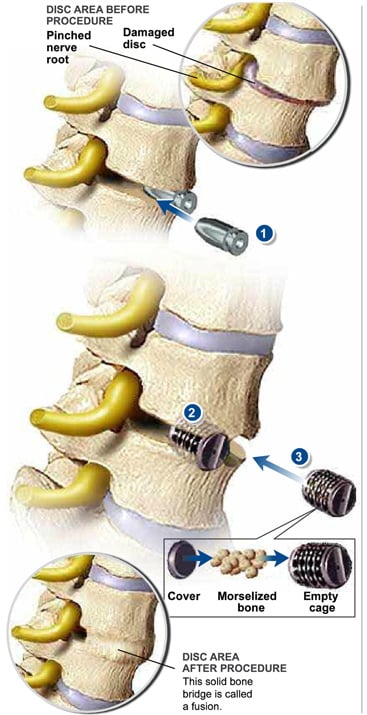
After removing the disc, the surgeon inserts temporary spacers into the empty space, as shown in illustration 1. This act realigns the vertebral bones and eliminates the pressure from your pinched nerve roots.
3. CREATING THE FIRST CHANNEL
The surgeon replaces the first spacer with a threaded metal cage (illustration 2). He or she creates a channel in the vertebral bones above and below the empty space to hold the cage. The surgeon screws in the cage, which is packed with bone graft, tightly into place.
4. CREATING THE SECOND CHANNEL
Your surgeon then removes the other temporary spacer and creates the channel for the second cage. He or she screws the second cage tightly into place, as shown in illustration 3.
5. RECOVERING FROM THE PROCEDURE
The morselized bone graft (illustration 3) eventually grows through and around the implants, forming a bone bridge that connects the vertebral bones of your spine. This solid bone bridge, shown in the bottom illustration, is called a fusion.
You must restrict your bending, twisting, lifting and driving for up to six weeks after surgery. You may start physical therapy after that.