This surgical procedure opens your spinal canal (the hollow space inside your spine) to relieve pressure on your spinal cord and nerves. To accomplish this, your surgeon increases the width, from side to side, of the roof of your spinal canal. Once secured in place, the procedure creates more room for your spinal cord, effectively addressing the problem.
If you’ve been diagnosed with myelopathy or spinal cord compression with symptoms such as weakness and/or numbness in your arm and hands, this procedure can reduce or eliminate your symptoms. The surgery is performed through an incision in the back of your neck.
OVERVIEW
This surgical procedure creates more space for your spinal cord and nerve roots, which relieves the painful pressure caused by spinal stenosis, a narrowing of the spinal canal. Some people are born with this ailment, but other causes include injuries, arthritis, tumors, herniated discs and bone spurs. The cervical laminaplasty procedure is described below. Ask your doctor about your risks and what to expect from your recovery.
 1. PREPARING FOR THE SURGERY
You are placed face down on the operating table. After the anesthesia has taken effect, your board-certified surgeon makes an incision on the back of your neck to access your spine. This incision leaves a minimal scar.
2. CUTTING THE VERTEBRA
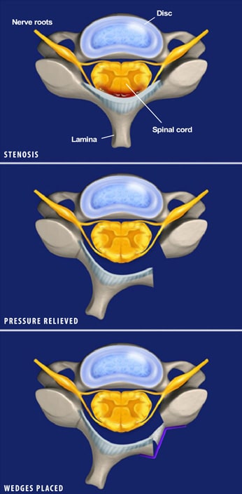
Your surgeon then moves aside the muscle to access your spine. Then on one side of the spine, the surgeon scores each lamina — the bony area that covers your spinal cord — to create a vertical groove down your spine. On the other side, the surgeon cuts through each lamina.
3. RELIEVING THE PRESSURE
The grooves on the first side act as hinges that allow the vertebrae to open like a door, as shown in the illustrations. This creates more space in the spinal canal, which relieves the pressure on the spinal cord and nerve roots. The surgeon inspects the spinal canal for bone spurs or other sources of compression and removes them.
4. PLACING THE WEDGES
To keep the more spacious bone passages open, your surgeon inserts small wedges made of bone graft material into each cut lamina. See the bottom illustration. The surgeon uses screws and metal implants, usually made of titanium, to secure the vertebrae and the bone wedges in place.
5. RECOVERING FROM THE PROCEDURE
After the surgeon closes and bandages your incision, they may place drains in the wound to prevent fluid buildup and encourage healing. The drains are removed after one or two days. You may be required to wear a soft cervical collar for up to two weeks, at which time you can return to deskwork. Full recovery usually requires three months, although you may need to do therapeutic exercise during that time.