Discography is a diagnostic procedure the back experts at the Southeastern Spine Institute (SSI) use to determine if any of your intervertebral discs are the primary cause of your back pain. Also called a discogram, this procedure is often followed with a Computed Tomography (CT) scan to confirm damage to the disc.
A discography procedure involves injecting a sterile liquid or contrasting dye into your discs to pressurize them. Healthy discs are flexible enough to withstand the pressure, but damaged discs cannot and will cause some pain.
 OVERVIEW
Discography helps your back specialist plan a course of treatment to alleviate your pain. The first step is to determine the source of your pain, which a Discography can do. The procedure is detailed below. Ask your back doctor about any risks that apply to you.
1. CONNECTING THE IV
For the procedure, you lie on a table equipped with a fluoroscope, either on your side or your stomach. Your doctor connects you to an intravenous (IV) line that administers medication to relax you. You must be awake enough during the procedure to tell the doctor what you feel.
2. INSERTING THE GUIDE NEEDLES
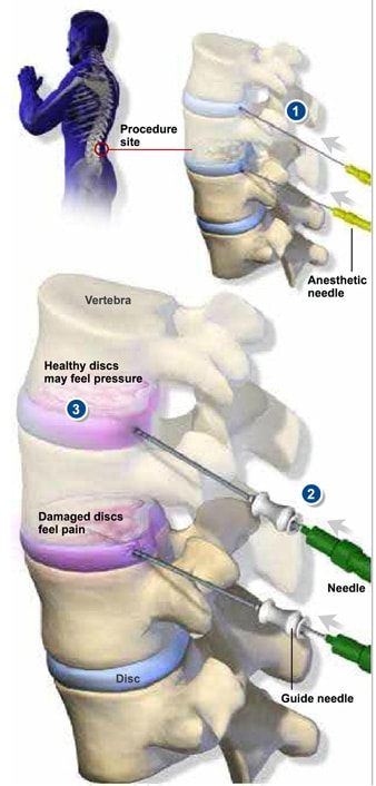
For each disc the doctor wants to test, he or she uses a local anesthetic to numb the skin above your spine and all the tissue down to the disc area. Illustration 1 shows this step.
Using the fluoroscope’s live X-ray images, your doctor identifies the location of the first disc to test. He or she inserts a guide needle through the anesthetized track to the outer edge of the disc. As shown in illustration 2, your doctor then inserts a smaller needle through the guide needle into the center of the disc. The back doctor may repeat this step several times if he or she needs to test more than one disc.
3. TESTING YOUR DISCS
Once your doctor has placed all the needles, he or she pressurizes the discs one at a time with injections of contrast dye. With each injection, you will feel either pressure or pain, as shown in illustration 3. If you feel pain, compare it to the pain you’ve been experiencing before your visit. If the pain feels the same, this may indicate a diseased disc.
4. REMOVING THE NEEDLES
After your doctor tests each disc, he or she uses the fluoroscope to take images of your discs. Then your doctor removes the needles. You may have to get a CT scan for additional images of the inside of your discs.
MORE INFORMATION
Contact us to learn more about the Discography procedure.
For more information about nerve blocks and epidurals, refer to SSI’s Block Suite.